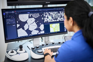
Scanning electron microscopy (SEM) in general can be used to explore intricate, ultrastructural 3D information with various methods collectively known as volume EM (vEM). Serial block-face SEM (SBF-SEM) is the vEM technology of choice for researchers who prefer easy sample preparation in combination with a highly automated imaging process enabling the acquisition of large volume datasets.
ZEISS Volutome is an end-to-end solution for serial block-face imaging from hardware to software including image processing, segmentation, and visualization. The ultramicrotome can be easily replaced by a conventional SEM stage, converting the 3D FE-SEM into a standard FE-SEM, making the system adaptable to a multi-purpose environment.
Saving time with automated cutting, image acquisition, and pre-processing
Imaging of biological structures with high resolution in large volumes can last for days. Therefore, SBF-SEM requires stable acquisition conditions over a long period of time. ZEISS Volutome allows highly automated and unattended cutting and imaging. During image acquisition, images are simultaneously pre-calculated for stitching and z-stack alignment. Results are at the user's fingertips in one click.
3D imaging in every way
ZEISS Volutome comes with the new high-speed, high-sensitivity detector that is specially designed for serial block-face imaging: ZEISS Volume BSD enables high-contrast images even at low acceleration voltages. In combination with the ZEISS patented Focal Charge Compensation, charge-prone samples can be easily imaged without compromises by charge neutralization at the block face. Furthermore, the built-in ZEISS Volutome stage facilitates the acquisition of large volume EM datasets.
Applications
Serial block-face imaging with ZEISS Volutome can be used to study cellular structures in any biological sample that has been prepared for electron microscopy. Neuroscience, cell biology and plant science are prominent research fields to mention or imaging of tissues in general.
From basic research in developmental, cellular, or molecular neuroscience to applied research in aging or neurodegenerative diseases, researchers across many neuroscience disciplines continue to pursue better understanding of neuronal connections and signaling pathways with serial block-face SEM.
In cell biology, high resolution imaging is necessary to visualize the ultrastructure of cells and cellular components, analyze morphology, and quantify structures of interest.
Serial block-face SEM reveals ultrastructural changes in plants induced by genetic mutations or suffering from drought, climate change or pollution. These factors translate into health and disease states in plants which impact crop yield, food production and, ultimately, human wellbeing.
Whether researchers work with tumors and biopsies, organ or tissue sections, organoids, embryos of model organisms and more, serial block-face imaging allows large sample tissue volumes to be imaged and analyzed within a broader 3D context.
One contact for serial block-face imaging
With years of experience in vEM involving serial block-facing imaging, ZEISS offers an end-to-end solution from hardware to software. ZEISS Volutome is based on the basic research technology of the Max-Planck Institute for Neurobiology in Martinsried, Germany. ZEISS is in close contact with the research community to provide solutions that are tailored to the users' needs.
About ZEISS
ZEISS is an internationally leading technology enterprise operating in the fields of optics and optoelectronics. In the previous fiscal year, the ZEISS Group generated annual revenue totaling 8.8 billion euros in its four segments Semiconductor Manufacturing Technology, Industrial Quality & Research, Medical Technology and Consumer Markets (status: 30 September 2022).
For its customers, ZEISS develops, produces and distributes highly innovative solutions for industrial metrology and quality assurance, microscopy solutions for the life sciences and materials research, and medical technology solutions for diagnostics and treatment in ophthalmology and microsurgery. The name ZEISS is also synonymous with the world's leading lithography optics, which are used by the chip industry to manufacture semiconductor components. There is global demand for trendsetting ZEISS brand products such as eyeglass lenses, camera lenses and binoculars.
With a portfolio aligned with future growth areas like digitalization, healthcare and Smart Production and a strong brand, ZEISS is shaping the future of technology and constantly advancing the world of optics and related fields with its solutions. The company's significant, sustainable investments in research and development lay the foundation for the success and continued expansion of ZEISS' technology and market leadership. ZEISS invests 13 percent of its revenue in research and development – this high level of expenditure has a long tradition at ZEISS and is also an investment in the future.
With over 40,000 employees, ZEISS is active globally in almost 50 countries with around 30 production sites, 60 sales and service companies and 27 research and development facilities (status: 31 March 2023). Founded in 1846 in Jena, the company is headquartered in Oberkochen, Germany. The Carl Zeiss Foundation, one of the largest foundations in Germany committed to the promotion of science, is the sole owner of the holding company, Carl Zeiss AG.
Further information at www.zeiss.com.
ZEISS Research Microscopy Solutions
ZEISS Research Microscopy Solutions is the leading provider of light, electron, X-ray microscope systems, correlative microscopy and software solutions leveraging AI technologies. The portfolio comprises of products and services for life sciences, materials and industrial research, as well as education and clinical routine applications. The unit is headquartered in Jena. Additional production and development sites are located in Germany, UK, USA and China. ZEISS Research Microscopy Solutions is part of the Industrial Quality & Research segment.
Further information at www.zeiss.com/microscopy
SOURCE ZEISS Research Microscopy Solutions


Share this article