ZEISS introduces new technologies advancing ophthalmic care
ZEISS presents AT LARA next generation EDoF IOL, new all-in-one OPMI LUMERA 700 surgical microscope, and CLARUS 500 Ultra-widefield imaging at ESCRS 2017
JENA, Germany and LISBON, Portugal, Oct. 7, 2017 /PRNewswire/ -- During the 35th congress of the European Society of Cataract and Refractive Surgeons (ESCRS) in Lisbon Portugal, the Medical Technology business group of ZEISS continues to expand its comprehensive portfolio of integrated ophthalmic diagnostic and surgical solutions with the introduction of new technologies that help doctors to more effectively and efficiently advance clinical care and provide superior outcomes for their patients.
For cataract, ZEISS launches AT LARA®, the next generation Extended Depth of Focus (EDoF) intraocular lens (IOL) providing patients the widest range of focus in this category. ZEISS also introduces the new all-in-one OPMI LUMERA® 700, which integrates a broad range of technologies into one microscope providing ophthalmic surgeons of all specialties a premium visualization and workflow platform. Additionally, ZEISS presents the new CLARUS™ 500 HD Ultra-widefield Fundus Imaging system which leverages legendary ZEISS precision optics to provide high resolution true color clarity across the entire retina, from the macula to the periphery.
"The new innovations being introduced by ZEISS at ESCRS 2017 underscore ZEISS' commitment to helping doctors achieve the best outcomes for each patient," says Dr. Ludwin Monz, President and CEO of Carl Zeiss Meditec. "At ZEISS, we are constantly striving to help doctors further improve their clinical performance and practice efficiency by perfecting solutions in new technology categories – such as ZEISS AT LARA in the new EDoF IOL category – and by continuing to integrate technologies – such as with the new all-in-one LUMERA surgical microscope."
ZEISS next generation EDoF IOL with widest range of focus
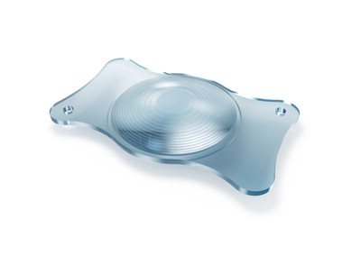
With the introduction of AT LARA® intraocular lens (IOL), ZEISS expands the company's market-leading IOL portfolio for cataract surgery into the new segment of Extended Depth of Focus (EDoF) IOLs. ZEISS AT LARA offers the widest range of focus within the EDoF segment with less visual side effects than multifocal IOLs.
With AT LARA from ZEISS, doctors can increase spectacle independence for a broader group of patients and address the growing need for improved intermediate vision performance, which is important for activities such as working at a computer. ZEISS AT LARA EDoF IOL enhances the range of vision for patients by creating an elongated focus range. Now doctors have a new option to provide superior visual outcomes for their cataract patients for whom multifocal IOLs might not be the optimal choice due to their patients' sensitivity to visual side effects, such as halo and glare at night.
"With AT LARA, ZEISS has expanded its comprehensive IOL portfolio to include a next generation EDoF lens which is showing superior visual outcomes and greater patient satisfaction than the first generation of EDoF IOLs," says James V. Mazzo, Global President Ophthalmic Devices at Carl Zeiss Meditec. "ZEISS AT LARA enables surgeons to expand their premium IOL offerings to treat a wider range of patients and provides them with a superior solution to meet each of their patients' needs."
The results of a pre-clinical trial1 have shown that ZEISS AT LARA next generation EDoF IOL provides a greater range of focus in comparison to first generation EDoF IOLs. The diffractive optical design of AT LARA creates an optical bridge effect to continuously extend the range of focus. The ZEISS IOL offers the widest range of focus among the EDoF segment providing patients with more spectacle independence for a wider range of activities. In clinical practice, ZEISS AT LARA has shown to induce less visual side effects than multifocal IOLs with patients experiencing optimized contrast sensitivity due to the aberration neutral aspheric design and the advanced chromatic correction. Additionally for the surgeon, the lens is 360° sharp edged to minimize PCO (posterior lens capsule opacification) and is pre-loaded for efficient and convenient surgical workflow.
Dr. Balasubramaniam Ilango, Medical Director of OPTIMAX clinics in the United Kingdom has been using the lens for the past seven months in his practice. "My patients are delighted and very happy with AT LARA EDoF lens. Their visual acuity is excellent over a wide range of distances, and so far they are not reporting any visual side effects."
ZEISS AT LARA: a new option for surgeons to provide individualized care
Dr. Florian Kretz, Fellow of the European Board of Ophthalmology (F.E.B.O.), and Chief Executive Officer and Lead Surgeon at the practice-clinic association of ophthalmologists, "Augenärzte Gerl, Kretz & Kollegen" in Germany, says, "AT LARA from ZEISS offers us an additional option for individualized patient care. It enhances the intermediate visual acuity and offers reduced optical phenomena with increased optical performance for distance and intermediate range." Dr. Kretz began implanting the lens for his cataract patients in March 2017. He continues, "All of these patients would choose a presbyopia-correcting lens again."
1 Data on file.
New OPMI LUMERA 700: premium ophthalmic surgical visualization and workflow technologies in one microscope
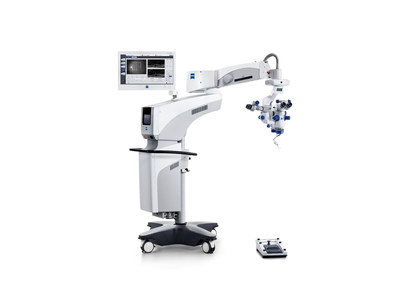
Continuing the successful OPMI LUMERA® ophthalmic surgical microscope product line, ZEISS introduces the new generation all-in-one OPMI LUMERA 700 at ESCRS 2017 providing ophthalmic surgeons of all specialties superior visualization and workflow technologies integrated into one microscope.
Improved IOL alignment tools and surgical assistance integrated into the OPMI LUMERA 700 address the growing demand for premium IOLs and optimal refractive correction. Intraoperative OCT imaging is supporting new ophthalmic surgical approaches and the treatment of more complex cases, leading to an increasing demand for even more advanced intraoperative imaging. The new ZEISS OPMI LUMERA 700 with its integrated functionalities addresses these trends and offers significant advances for cataract, cornea, glaucoma, and retina surgeons as well as teaching physicians, eliminating the need for separate devices for different specialties.
"Doctors no longer have to decide on either markerless toric IOL alignment or intraoperative OCT. They can now have both in one integrated solution," says James V. Mazzo, Global President Ophthalmic Devices at Carl Zeiss Meditec. "Surgeons across ophthalmic specialties will benefit from the substantial advancements in intraoperative OCT image quality, the leaner workflows and deeper integration. For example, our ZEISS Cataract Suite is even better and more efficient because of improvements in one of its major components, the OPMI LUMERA 700 microscope."
Due to enhanced algorithms and the streamlined workflow of the computer-guided surgical assistance of ZEISS Cataract Suite markerless, surgeons need to take significantly less steps and can align toric IOLs more precisely. "This is yet another process improvement of the proven ZEISS Cataract Suite workflow," Mazzo adds. Additionally a comprehensive "cockpit" provides surgeons relevant information during surgery – in the eyepiece, on the screen and in the video function. Phaco parameters and IOL data shown in the cockpit, for example, help eliminate the need for cataract surgeons to look up from their surgical view.
Retina, cornea and glaucoma surgeons benefit from improved intraoperative OCT image quality that is now closer to diagnostic OCT image quality. Surgeons can more easily correlate intraoperative OCT images delivered by the new OPMI LUMERA 700 to pre-operative retina and anterior segment diagnostic OCT images from the ZEISS CIRRUS™ OCT family. Thus, surgeons can see with more clarity and compare the details of critical structures and pathologies below the surface of the surgical field.
The integration of a broad range of ophthalmic surgical visualization technologies into one premium microscope platform supports hospitals and clinics that serve several specialties in their clinic with a cost effective capital investment solution. The new OPMI LUMERA 700 also comes with a 3D visualization option serving the needs of teaching institutions.
ZEISS CLARUS 500 Ultra-widefield Fundus Imaging: True Color clarity from the macula to periphery
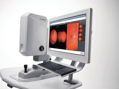
During ESCRS 2017, ZEISS presents CLARUS™ 500: the first fundus imaging system combining True Color with exceptional clarity within an ultra-widefield view. Early signs of eye disease can often be subtle and can occur in the far periphery of the retina. Now with ZEISS CLARUS 500 Ultra-widefield system, practitioners can obtain a better view of the entire fundus.
CLARUS 500 Ultra-widefield Imaging system leverages legendary ZEISS precision optics to provide high resolution retinal imaging down to 7 microns across the entire retina -- from the macula to the far periphery. CLARUS 500 produces exceptional images in True Color that closely resemble the coloration of the retina as seen through direct observation during clinical examination. Color accuracy is important in the diagnosis, documentation and management of ocular diseases; ensuring confidence when evaluating optic disc, nevi, and lesions in which subtle color differences may lead to a change in diagnosis and management. In addition to capturing natural-looking images of the fundus, the True Color images of ZEISS CLARUS can be separated into red, green, and blue channel images, which can enhance the visual contrast of details in certain layers of the retina.
With a single capture, ZEISS CLARUS 500 produces a 133-degree HD widefield image. HD widefield images are automatically merged to achieve a 200-degree ultra-widefield of view. Exceptional clarity from the posterior pole to the periphery, along with an intuitive review software, allow clinicians to track subtle changes in pathology, thereby helping with disease and patient management. Another advantage of the CLARUS 500 is its ability for peripheral imaging while still maintaining the ability to zoom into the retina without losing resolution.
"Traditional fundus imaging systems have been the gold standard for macular disease diagnosis and optic nerve evaluation for many years," says Jim Mazzo, Global President Ophthalmic Devices at Carl Zeiss Meditec. "Now, Ultra-widefield is starting to change this. Clinicians are finding that by imaging a larger area of the retina, they have the possibility of uncovering more pathology, aiding in earlier disease diagnosis and better patient management. With ZEISS CLARUS, they can better manage a broader range of patients with one fundus imaging system," Mazzo continues.
Simple, stable and intuitive, the technology allows clinicians to easily review and compare high-quality images captured during a single exam while providing annotation and caliper measurement tools that allow in-depth analysis of eye health. CLARUS 500 has also been designed to optimize each patient's experience. By bringing the optics to the patient, and with Live IR Preview for technicians to easily ensure optimal patient alignment, ZEISS CLARUS 500 helps create a comfortable, satisfying patient experience that provides images free of obstructions, such as lids and lashes, and requires fewer recaptures.
CLARUS works with ZEISS FORUM® and Retina Workplace for review with other ophthalmic images and exam data for efficient multi-modality analysis.
During the 2017 ESCRS in Lisbon, renowned experts will be sharing their experiences with the latest technologies from ZEISS, and attendees can experience first-hand ZEISS' integrated ophthalmic diagnostic and surgical solutions at the ZEISS booth P262 from October 7-11, 2017 at the FIL Feira Internacional de Lisboa.
For more information ZEISS' scientific and educational program and events at ESCRS:
www.zeiss.com/escrs
Photo - https://mma.prnewswire.com/media/569547/ZEISS_AT_LARA_EDoF_IOL.jpg
Photo - https://mma.prnewswire.com/media/569543/ZEISS_CLARUS_500_1.jpg
Photo - https://mma.prnewswire.com/media/569545/ZEISS_AT_LARA_EDoF_IOL_2.jpg
Photo - https://mma.prnewswire.com/media/569546/LUMERA_700_NEW.jpg
Logo - https://mma.prnewswire.com/media/569544/ZEISS_Brand_RGB.jpg


Share this article